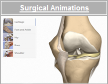
Bones
The knee joint is made up of four bones:
- The femur is the bone of
the thigh. It is the largest bone in the body.
- The tibia is the large
bone in the lower leg. The femur sits on the tibia.
- The fibula is the smaller
bone in the lower leg. It serves as an attachment point for muscles
and the lateral collateral ligament.
- The patella is also known
as the "kneecap". It is located in front of the femur and tibia. As
the knee moves, the patella slides within a groove on the femur.
Back to top
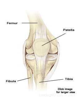
Ligaments
Four major ligaments connect the bones of
the upper and lower leg. Ligaments are strong bundles of fibers that stabilize
the joint, guide joint motion, and prevent excessive motion.
-
Anterior
Cruciate Ligament (ACL) and Posterior Cruciate Ligament (PCL)
The cruciates are the two major ligaments inside the knee joint. The
name "cruciate" means "cross" and comes from the fact that these two
ligaments cross each other as they attach to the femur and the tibia.
-
Medial
Collateral Ligament (MCL) and Lateral Collateral Ligament (LCL)
The collateral ligaments are the ligaments on either side of the knee
joint. The MCL is on the inner side of the knee and the LCL is on
the outer side of the knee.
Back
to top
Muscles
and Tendons
 Two
sets of muscles cross the knee joint to make it move. Two
sets of muscles cross the knee joint to make it move.
-
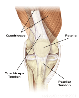
The quadriceps (sometimes referred to as "quads") are
four muscles in the front of the thigh that straighten the knee .
-
The hamstrings (sometimes referred to as "hams") are
the muscles in the back of the thigh that work together to bend the
knee.
Tendons are the connective
structures that attach muscle to bones. Ligaments connect bone to bone.
The four quadriceps come together to form one tendon called the quadriceps
tendon. This tendon surrounds the patella and is called the patellar
tendon as it attaches the muscles to the tibia.
Back
to top
Cartilage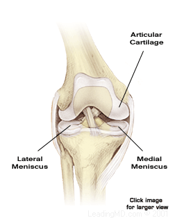
There are two types of cartilage within the
knee:
Articular Cartilage - The ends of each
bone are covered with this smooth substance. Articular cartilage serves
two purposes:
it minimizes friction and wear of the bone surfaces.
it spreads the loads that are applied to a joint.
Meniscus - There are two C-shaped
wedges called menisci (plural). The medial meniscus and the lateral
meniscus are cushions between the femur and the tibia. These rubber-like
shock absorbers improve the fit of the two bones. The menisci are the
parts of the knee damaged when someone is said to have "torn cartilage."
© 2015 by LeadingMD.com All rights reserved.
Disclaimer
|